


OcomeComyCosis is a type of fungal lesion, which affects exclusively on the nail.To prevent the spread of infection, you should know what the nail fungus looks like, because the therapeutic effect will be achieved faster if the treatment has begun at an early stage of infection.The most common disease occurs in older people due to low immunity, but the risk of disease to refer to each person.There is no unique classification of fungal nail damage;In medical practice, it is common to distinguish them at the location of the localization and depth on which they penetrate.The infection is also grouped depending on the type of pathogen.
Types of pathogens and primary characters
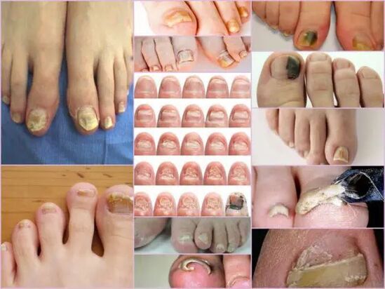
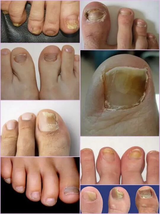
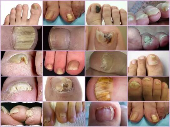
The symptoms of nail fungus in the initial phase are easily determined directly under the term of nail.This sign is the most upbound, because onychomicosis is always manifested in the form of colored surface changes, beds of bed, peeling and any external changes.The latter is expressed by roughness, the formation of grooves and cracks, as well as points intertwined and general breach of the nail.
The primary sign of a healthy nail is pink and transparency.Thosehomicosis at any stage is characterized by a nail blur and color change in yellow, brown or greenish-black.In the advanced phase of surfaces, it can gain black shade along the background of almost complete destruction.
External signs of fungi infection depend on the type of pathogen that infected.In medical practice, the following possible lesions differ:
- Candida Infection to fungi, which is expressed by discharging the nail directly at the foot of the box.The candidiasis of the roller around the nail will be characteristic of the candidate.This version of complications may have either a bacterial source in the form of streptococci or staphylococci or expressed in the form of medium or psyrian plaques;
- Dermatophytes type Trichophyton Rubrum.In this case, the penetration of infection occurs directly from the free edge of the nail.The first symptoms of such pathogen phenomena are a yellow place on the surface.In the field of neoplasm, the nails fall apart, and the place itself is increasing.The usual place of localization of neoplasm is along the slab, in parallel with nail rollers, in this case the infection is called Distal-Side.There is another form of defeat from this pathogen - distal, in which the place appears on the part removed from the hole, mainly in the middle of the free edge;
Important!
This type of fungus that most often occurs on the legs of their legs, gradually rotates the nails in a loose yellowish mass.In the absence of proper treatment, the disease is poured into hypercheralatosis.The nail plate is completely destroyed due to the spread of the infection around the rim.
- Dermatophytes like a trichophyton mentagrophyte.Thosehomicosis with such exciting is most often based on thumb nails, less often than small fingers.Infection with fungius of this type requires therapy not just nails, but also legs due to the rapid spread of pathogen.Symptomatic disease can be like Leikonichia, a disease that is common in medical practice.Primary signs are white places that appear on various nail parts, neoplasms are characterized by non-standard shape and various sizes.It is easy to distinguish between Leakonichia fungus - in the latter case, the points are the accumulation of air, which is not observed with fungal impairment;
- Mold fungus.This damage option is found significantly less often than candidals or dermatophytic form.The main sign only such infections - the surface of the slab acquires a dark, almost black shade.It is not fully infected all the finger, but only part of the nail plate.The first signs of fungus nails on the legs of this type are sharp color changes.Thoseomicosis can be developed in the form of a longitudinal strip of black or dark green color in color from the background of the rest of the pink part of the nail.
Diagnostics according to the type of change
It is not difficult to notice the nail fungal on their feet even with the primary phases of the infection, as the infection of this type is obviously manifested quite active from the first day of the lesion.Instead of common transparent nails pale pink pink in the patient, significant surface deformations and general changes are observed.There is a matte tupa yellowish shade on the convenient area, which seems mostly on thumbs.The type of fungus and the degree of damage are determining the factor.
In the first phase, fungal feet damage on your feet is the look of a small fare.There will be a characteristic thickening of the board and keratysis of the bed under the nail.This phase is accompanied by such a phenomenon as a partial detachment and discharge of nail plate, which serves as a source of infection for healthy people.
Despite the active thickening of the plate, its constant grinding can be observed, regardless of current factors.The features of each phase and symptoms of the fungus on the legs depend on the type of pathogen.
Depending on changes that occur with nail plate, three options for onychomicosis are different:
- Hypertrophic;
- Normotroph;
- Atrophic.
In the first case, in the shade of nail plates, in the first case, its destruction along the edges, as well as the deformation of the surface of the plate.The nail is so thickened to cause discomfort and painful sensations when walking.The normotrophic micosa nails on their feet is characterized by the presence of a healthy glow, but the board itself acquires some cravings, stains form on it.With the atrophic type of damage, brown and gray focal forms on the surface of the nail.Thanks to them, it is possible to determine the location of the pathogen exactly.
Important!
Attrophic or onycholytic type of nail fungus is characterized by thinning the nail plate, not its growth, as in other cases.The areas where pathogen is localized usually separate.The lack of proper treatment leads to an advanced phase - completely rejection of the nail plate.
Localization classification
Another sign by which you can separate fungal damage on your toes, depends directly on the hotspot site on the nail plate.This includes the depth of pathogen, which in turn allows you to determine the approximate duration of the upcoming therapy and a chance for a speedy recovery.
Fungal diseases Nail legs at the location of localizations are classified into the following groups:
- OnihyComyCosis is a type of white surface - the appearance of many whitish stains on the surface of the nail plate.A fungal infection leads to separation of the skin in places of appearance of stains where there is active emptying of the scales.Advanced phase leads to complete destruction and deduction of plates;
- Distal - develops on the free edge of the nail.Color change is observed first in the small area of the plate, after which it is an active expansion of its limits.Lesia is characterized by yellowish gray or brown strings, as well as rough uneven surfaces and gradual peeling;
- Side ondychomicosis has stages of development similar to distal, but is localized exclusively on the sides of the nail plate;
- Total infection - complete infection of the entire surface;
- Proximal onnikhycosis.This disease begins his activity from the cuticle, and then the fungus is rapidly affected by the nail, and the process itself begins with the appearance of a small white place near the lapse of the perikologue roller.
The most common forms of micosome nails are side and distal, which usually cause dermatophytes.Such forms of damage as proximal and white can act as a secondary disease that monitors the disease of the immune system, for example, VICH.Total nail damage with fungi should be considered an advanced phase of the development of any fungus under the nail.
Features of the fungus under the microscope
Despite the presence of an impressive number of the number of micromicosis, other diseases related to skin problems and not infectious nature are accepted for fungal damage to nails.You can specify the correct diagnosis and type of pathogen only in the lab under the microscope, for which the hospital in the hospital is to scrape the biological material from the affected areas.
The resulting biomaterial is placed in advance in an alkaline environment, after which multiple growth is done.Study Fungus for nail under the microscope allows you to see the active pathogen, from which the external form determines its type, distribution scale and approximate degree of damage.You can predict approximate colony growth in nutrients, which allows not only to be precise diagnosis, but also to determine the restriction of infection.
Important!
Since it is possible to determine the presence of onnicmicosis only in the laboratory, you should not postpone the visit to the doctor at the slightest doubt.Fungus nails are developing rapidly, and the waste of time increases therapy.
What is an alarming signal?
Mushroom nails on their feet manifests certain symptoms that are partly similar to some skin diseases.The following signs are more likely to indicate an onychomitoleafly lesion that requires medical intervention:
- The occurrence of yellow spots on the plate, its deformation, changing the structure, which was not observed earlier;
- Blackberry discoloration, transparency loss, some photographs of the nail nails show a shade near black, which is characteristic of mold pathogens;
- Thickening and keratinization of nail plates or vice versa too sharp thinning;
- Rejection of the nail out of bed, separation of scales and crumbs;
- Rolling down the roller, hangs above the plate, the release of the liquid;
- Nails affected by fungus appear inflamed, regardless of the type of pathogen.
All these symptoms indicate a high risk of infection.Some external symptoms of changes in nail plate structure can be the result of other diseases.With an increased better, amount of calcium and iron in the body should increase, a gomolic essential infection of the body is increased, with pure white stripes and points, is possible a lack of copper or zinc.Despite the fact that in the photograph, the nail fungus seems like focal damage to the color change with a color closer to yellow, it does not always indicate the infection.Yellowness may indicate different problems with the stomach and liver.
It is not difficult to recognize onychomikosu on the fingers external signs, despite the wide classification of the disease.But you can specify the exact type of pathogen and damage phase can only be in the laboratory.